Year 2025 / Volume 36 / Number 1

Clinical Cases

Epidermoid carcinoma associated to onichocryptosis: a case report

Rev. Esp. Pod. 2025; 36(1): 48-53 / DOI: 10.20986/revesppod.2025.1729/2025

Antonio Javier González Fernández, Lucía Trincado Villa, Javier Pascual Huerta, Alicia Gavillero Martín


ABSTRACT

Primary malignant tumors located in the nail or subungual area are rare, with squamous cell carcinoma or squamous cell carcinoma being the most common of all of them. The present work describes the progression of a clinical case of a patient with initial diagnosis of onychocryptosis, with a torpid evolution of the lesion and who after various conservative and surgical treatments ended with a diagnosis of squamous cell carcinoma and complete amputation of the first finger. The article shows the entire progression of the clinical picture from its beginning, emphasizing the importance of a high index of suspicion on the part of the clinician and the need for diagnosis through pathological anatomy in the event of an abnormal evolution of the lesion.


RESUMEN

Los tumores malignos primarios de localización ungueal o subungueal son poco frecuentes, y el carcinoma epidermoide o carcinoma escamoso es el más común de todos ellos. El presente trabajo describe la progresión de un caso clínico de una paciente con diagnóstico inicial de onicocriptosis, con una evolución tórpida de la lesión la cual, tras diversos tratamientos conservadores y quirúrgicos, acabó con un diagnóstico de carcinoma epidermoide y amputación completa del primer dedo. En el artículo, se muestra toda la progresión del cuadro clínico desde su comienzo, enfatizando en la importancia de un alto índice de sospecha por parte del clínico y de la necesidad del diagnóstico mediante anatomía patológica ante una evolución anormal de la lesión.


Complete Article


10.20986/revesppod.2025.1729/2025
CLINICAL CASE

Epidermoid carcinoma associated to onichocryptosis: a case report
Carcinoma epidermoide asociado a onicocriptosis: caso clínico

Antonio Javier González Fernández1
Lucía Trincado Villa1, Javier Pascual Huerta1
Alicia Gavillero Martín2

1Clínica del Pie Elcano. Bilbao, España.
2Departamento de Podología General Universidad Católica de Valencia “San Vicente Mártir”. Valencia, España

Abstract

Primary malignant tumors located in the nail or subungual area are rare, with squamous cell carcinoma or squamous cell carcinoma being the most common of all of them. The present work describes the progression of a clinical case of a patient with initial diagnosis of onychocryptosis, with a torpid evolution of the lesion and who after various conservative and surgical treatments ended with a diagnosis of squamous cell carcinoma and complete amputation of the first finger. The article shows the entire progression of the clinical picture from its beginning, emphasizing the importance of a high index of suspicion on the part of the clinician and the need for diagnosis through pathological anatomy in the event of an abnormal evolution of the lesion.

Keywords: Foot, nail, onychocrytosis, epidermoid carcinoma, squamous carcinoma, Winograd, amputation

Resumen

Los tumores malignos primarios de localización ungueal o subungueal son poco frecuentes, y el carcinoma epidermoide o carcinoma escamoso es el más común de todos ellos. El presente trabajo describe la progresión de un caso clínico de una paciente con diagnóstico inicial de onicocriptosis, con una evolución tórpida de la lesión la cual, tras diversos tratamientos conservadores y quirúrgicos, acabó con un diagnóstico de carcinoma epidermoide y amputación completa del primer dedo. En el artículo, se muestra toda la progresión del cuadro clínico desde su comienzo, enfatizando en la importancia de un alto índice de sospecha por parte del clínico y de la necesidad del diagnóstico mediante anatomía patológica ante una evolución anormal de la lesión.

Palabras clave: Pie, uña, onicocriptosis, carcinoma epidermoide, carcinoma escamoso, Winograd, amputación

Correspondence
Antonio Javier González Fernández
antgonfer94@gmail.com

Received: 09-03-2025
Accepted: 02-05-2025

Introduction

Primary malignant tumors of subungual localization are rare. Of these, squamous cell carcinoma (SCC) is the most prevalent in the foot(1,2). The most affected patients are between 50 and 60 years old, affecting men more than women, in a ratio of 3:12. The most common presentation is in the proximal and lateral nail folds and hyponychium and is characterized by erythema, paronychia, ulceration, hyperkeratosis, verrucous papules and plaques, subungual mass, onycholysis, and suppuration. These non-specific clinical characteristics mean that it can be easily confused with benign inflammatory processes such as paronychia, pyogenic granuloma, or a common wart, which often delays its initial diagnosis. When it affects the nail matrix, it can present with longitudinal erythronychia, melanonychia, and nail destruction(3). The diagnosis of these types of lesions is only possible through a biopsy and subsequent histopathological study. Treatment of these tumors involves complete excision, which sometimes means amputation of the distal phalanx of the affected toe(1,3,4,5,6,7).
This article illustrates the case report of a patient with a past medical history of onychocryptosis in the first toe of the foot with a torpid progression over time despite conservative and surgical treatment with several interventions, whose final diagnosis was squamous cell carcinoma. The objective is to present these types of cases to the podiatric community, highlighting the importance of performing a histopathological study in suspected cases or in those where the lesion evolves abnormally with conventional treatment. The article has been prepared following the CARE (CAse REport Statement) guidelines for the reporting and development of clinical cases in scientific journals(8).

Case report

A 50-year-old woman presented with a several-month history of pain and deformity in the first toe of her left foot in the context of several previous interventions on the first toe for medial border onychocryptosis. Relevant medical history includes no known drug allergies, smoker of 30 cigarettes/day, hypercholesterolemia, chronic diarrhea due to collagenous colitis, endometriosis, and depressive syndrome under psychiatric follow-up. Currently on treatment with: budesonide 3 mg (2-0-0), escitalopram 20 mg (1-0-0) and lormetazepam 2 mg (0-0-1), medroxyprogesterone 5 mg (1-0-0), calcium/vitamin D 1.5 g/400 IU (1-0-0), progesterone 100 mg (2-2-0).
The patient reported a 7-month history onychocryptosis (September 2022) on the medial border of the first toe of the left foot, with presence of ungual granulation tissue on said border, causing her pain and digital inflammation (Figure 1). The patient received treatment from her podiatrist consisting of chiropody and canal cleaning, and then daily dressings with mupirocin were prescribed for the lesion. The lesion showed poor progression in the following days with increased pain and tenderness to the touch. Treatment was modified to amoxicilin/clavulanic acid 875/125 mg, 1/8 hours along with the use of oral analgesics (Figure 2). The patient continued with poor symptomatic evolution and attended her health center where an X-ray was performed, ruling out the presence of deep infection (osteomyelitis).

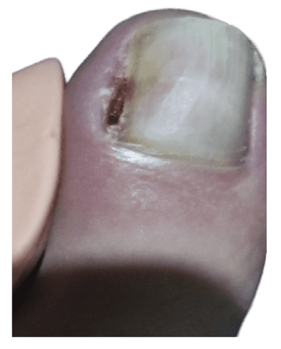
Figure 1. Initial process of onicochryptosis and reactive granuloma.

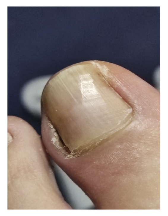
Figure 2. Clinical progressión after antibiotic and analgesic treatment.

After worsening of the clinical picture, on November 14th, 2022, a partial removal of the lateral nail plate of the left foot was performed at her health center along with curettage, removing hyperkeratotic and granulation tissue. The procedure was repeated 48 hours later (November 16th, 2022) because not all the lesion had been removed (Figure 3), changing the antibiotic treatment to anaclosil 500 mg 1/6 hours and then to ciprofloxacin 500 mg 1/12 hours during the following weeks.
The patient initially showed improvement in pain, although it never completely subsided (Figure 4). She reported recurrence of the nail lesion, which led her to seek a second opinion 6 weeks after the intervention with another podiatrist. This latter podiatrist, on December 27th (Figure 5), performed a bacterial culture of the lesion, which turned out negative, and also evaluated the lesion by ultrasound (without report). On January 13th, the patient underwent surgery using the phenol-alcohol technique on the lateral border of the first toe of the left foot (Figure 6).

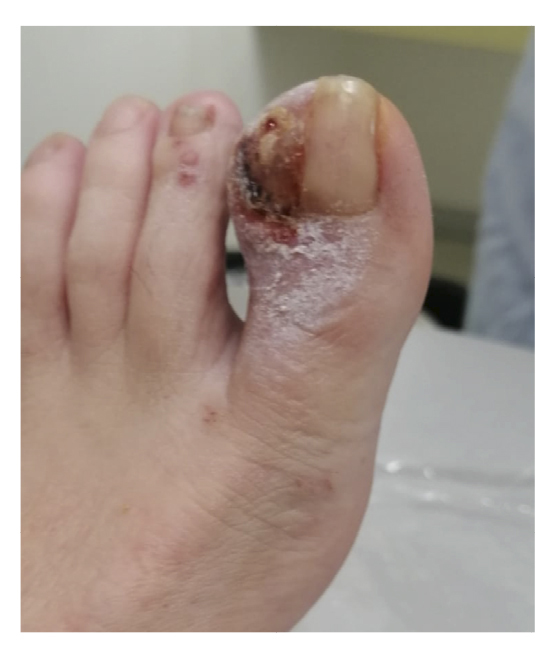
Figure 3. 8th week. Partial resection of hyperplasic tissue performed in her medical center.

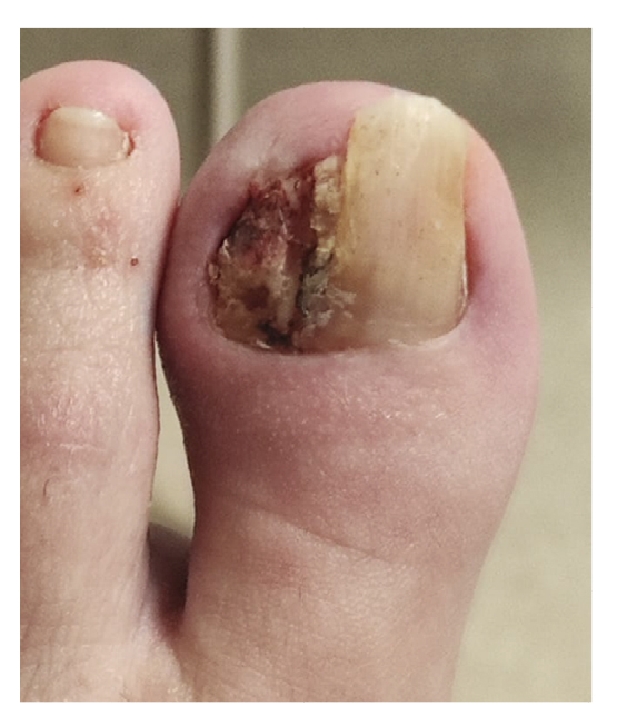
Figure 4. 9th-14th weeks. Recurrence of the lesión after partial resection.

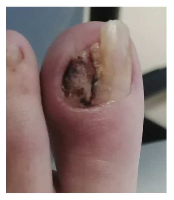
Figure 5. 16th week. Negative microbiological culture and sonograhpic assessment (not available).

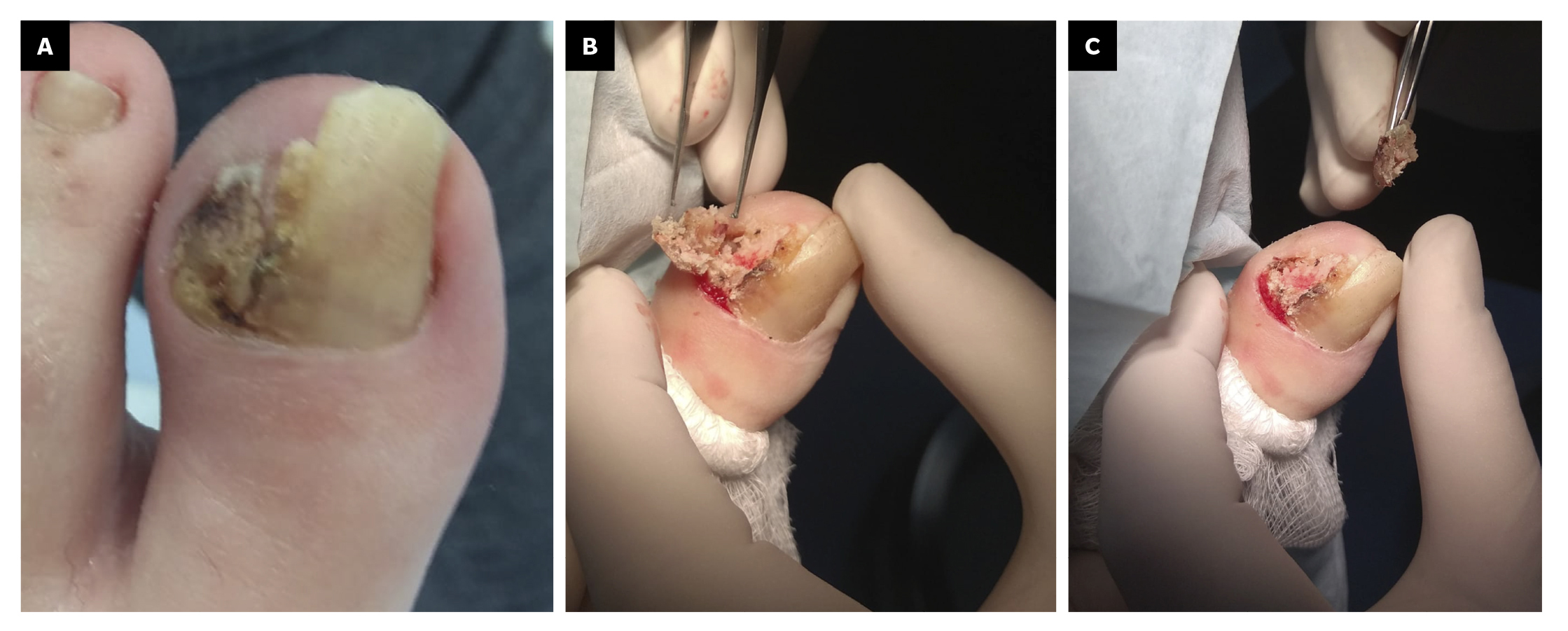
Figure 6. 18th week. Second intervention by means of phenol-alcohol partial matricectomy.

Within 3 weeks after the third intervention, the patient reported recurrence of the lesion and painful symptoms (Figure 7). The patient decided to attend the traumatology department (February 2023) where an MRI (T2FS and DPFATSAT) was requested. It showed an occupying mass in the lateral third of the first toe with well-defined borders and bone involvement (Figure 8) with the following report: “At the level of the distal phalanx of the 1st toe in the nail region, there appears to be a solution of skin continuity with a heterogeneous, a dense 15 mm x 14 mm collection in the lateral region of the parapharyngeal soft tissues, remodeling the bone and coming into contact with the flexor tendon, which could be consistent with a postoperative abscess/phlegmonous collection. Some bone edema in the distal phalanx is suspected, without completely ruling out incipient osteomyelitis. To be correlated with medical history and surgical protocol performed.”


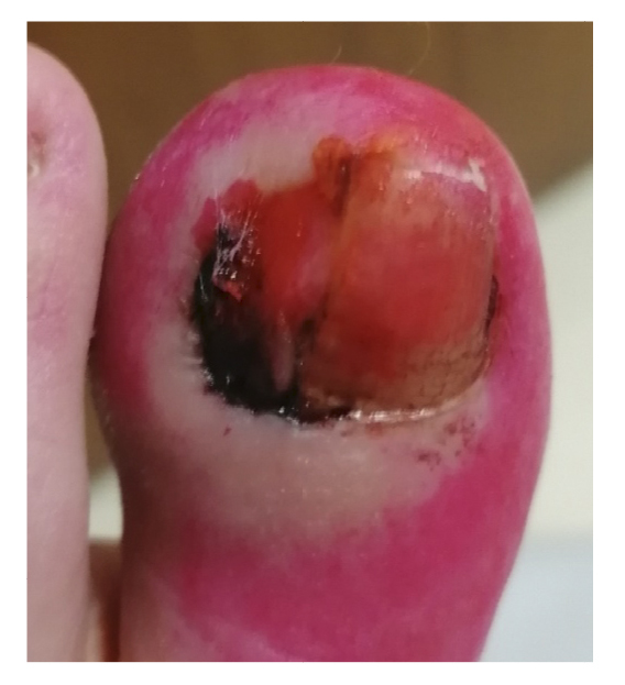
Figure 7. 21th week. Recurrence of lesion after phenol-alcohol surgery.


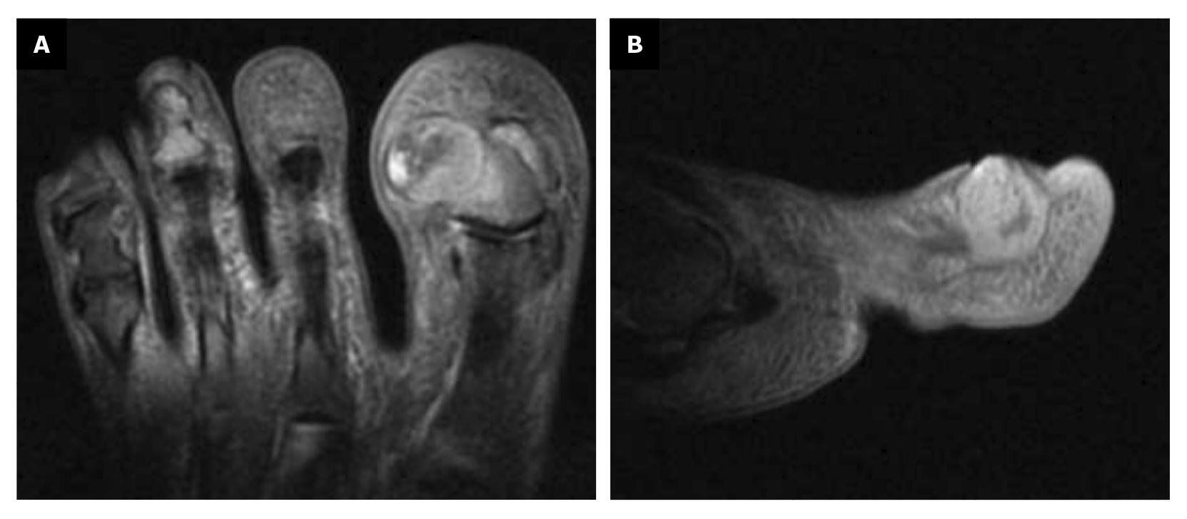
Figure 8. Space ocuping mass with bone involvement. A: transverse plane; B: saggital plane.


The Traumatology Service referred the patient to us, and she attended our center on February 27th, 2023. On March 1st, 2023, we decided to perform surgery with excisional biopsy of the lesion, with 5 mm safety margins around its contour. The lesion was completely removed, and given the bone involvement of the lesion, partial resection of the lateral border of the distal phalanx of the 1st toe was performed. Both samples (soft tissue and bone) were sent to pathology separately. Finally, the incision was closed with approximation sutures (Nylon 4/0) in the proximal and distal areas; since the primary defect was too large, the central area of the incision was left open to close by secondary intention with the use of hydrofiber (Figure 9).


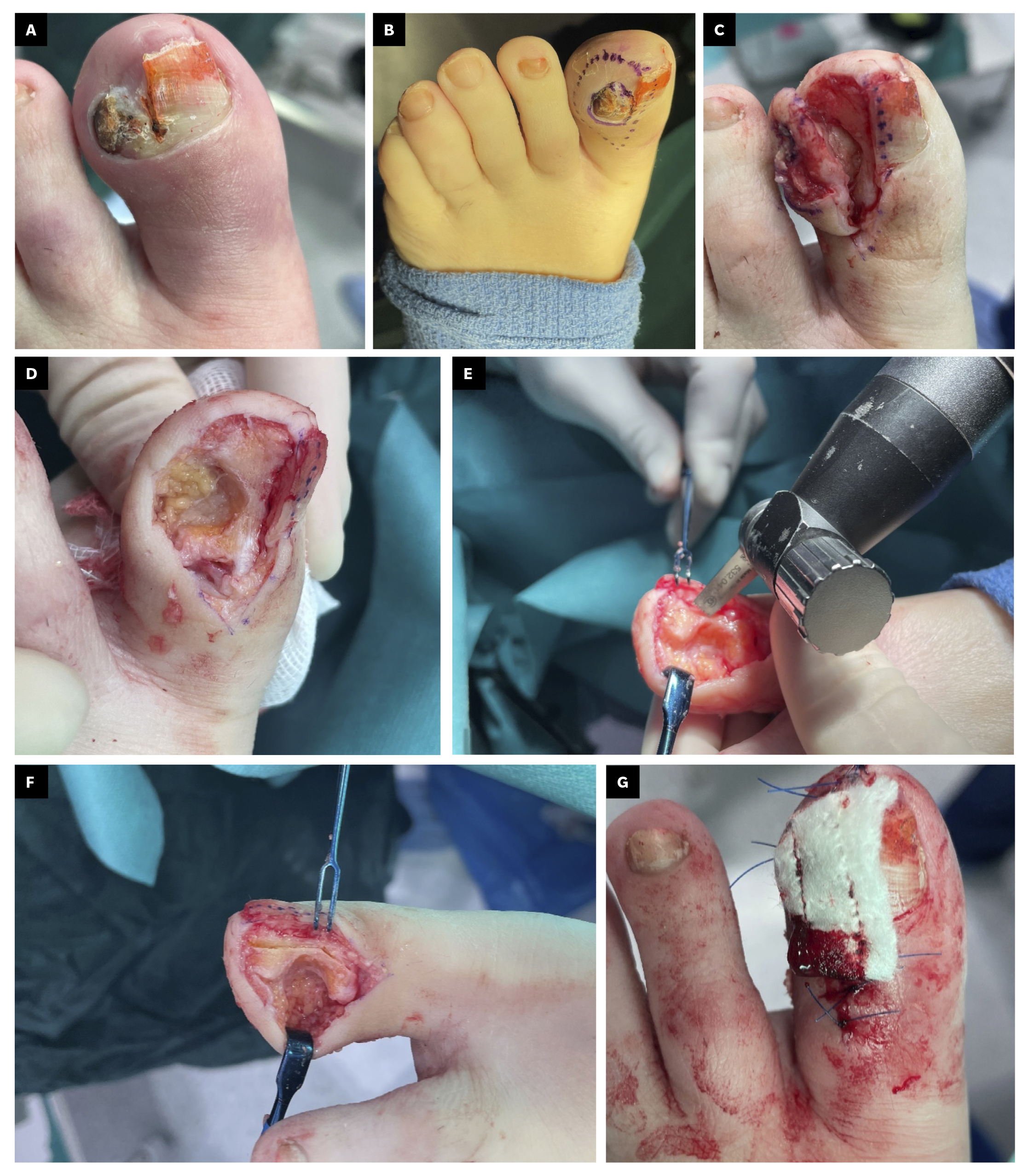
Figure 9. A: 26th week; clinical aspect of the lesion; B-F: secuence of tumor excision with security margins and partial resection of distal phalanx.

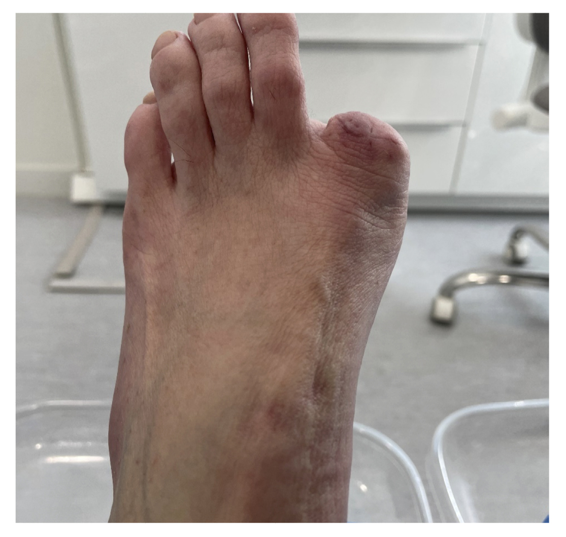
Figure 10. 30th week with digital amputation.

The histopathological study of the samples described for the first sample: “Skin with a hyperkeratotic tumor composed of squamous cells with discrete atypia forming an expansive border with some cords and nests infiltrating the dermis. Lateral surgical margins free and deep margin affected. DIAGNOSIS: Well-differentiated infiltrating squamous cell carcinoma affecting the deep margin,” and for the second sample: “Bone tissue with bone trabeculae and adipose tissue between them without alterations. Focally, two small nests of well-differentiated squamous cell carcinoma are observed. DIAGNOSIS: Bone tissue with focal infiltration by well-differentiated squamous cell carcinoma.”
5 days postoperatively, the patient’s first dressing change was performed, which showed a good appearance and no local infection. The patient was explained the result of pathology and referred to the oncology service of her hospital, and from there to the dermatology and plastic surgery service where digital amputation was performed (Figure 10). Subsequent follow-up was conducted by the Oncology service for the next 6 months. After different tests, it was determined that no other treatment was needed. Figure 11 shows the timeline of the clinical case.


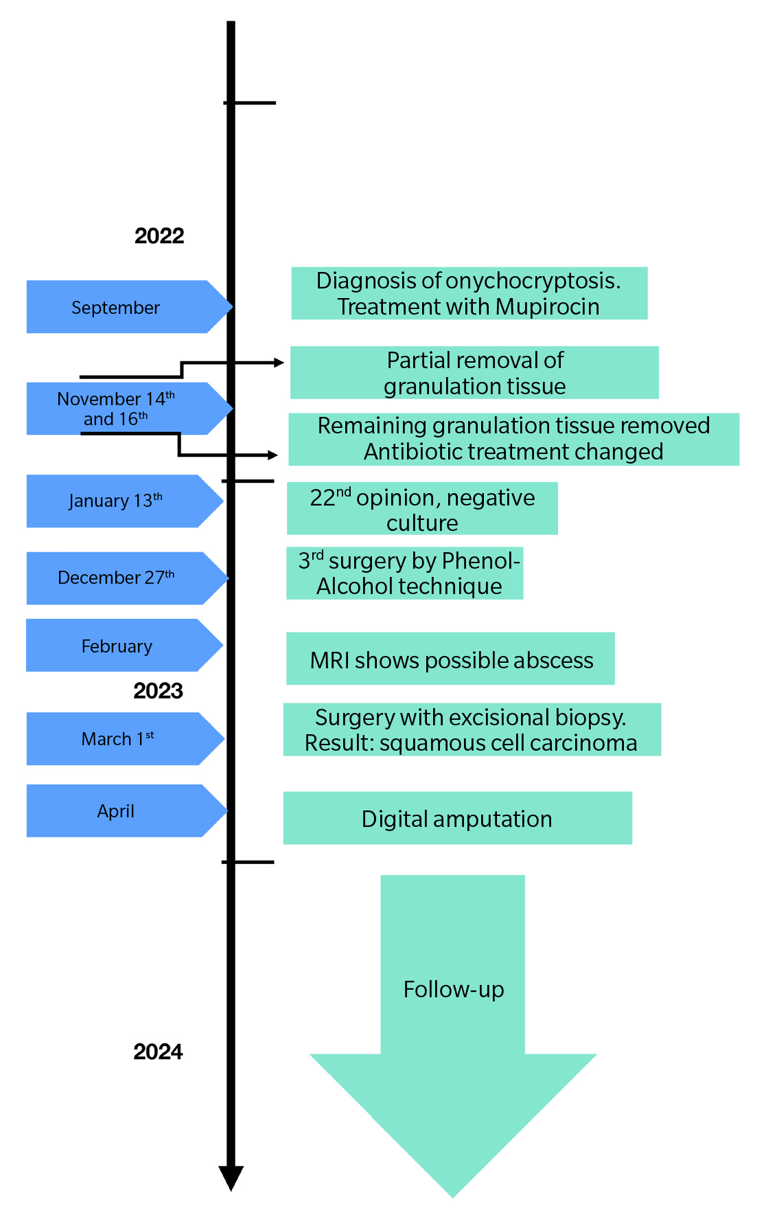
Figure 11. Case timeline.

Discussion

This article presents an atypical case of onychocryptosis, which ultimately led to a diagnosis of squamous cell carcinoma and resulted in complete amputation of the first toe. The objective is to introduce this pathology, although rare, to the podiatric community. Subungual squamous cell carcinoma (SCC) was first described by Velpeau in 1850. It is a neoplasm with low aggressive potential. It has a tendency to affect bone (described in up to 66% of cases) as in the case we presented. However, it rarely metastasizes(1,2).It frequently presents in men between 50 and 70 years old. Although it is more common in fingers, it can also occur in toes(4).. Up to 75% of cases are associated with human papillomavirus, especially serotypes 16, 18, 35, 56. In the feet, risk factors are thought to be: ulcers, chronic granulomas, trauma and chronic inflammation, exposure to tobacco smoke or solar radiation(2,3,4,5).
The clinical presentation depends on the location of the pathological process within the nail unit. When it affects the proximal or lateral nail fold or the hyponychium, it can present as erythema, verrucous papules, onycholysis, or suppuration. If it affects the nail matrix, it presents with longitudinal erythronychia and frequently nail destruction. If it affects the nail bed, it can present with onycholysis and suppuration, along with red papules and nodules(2,3).
It is a progressive and invasive tumor, typically presenting with a delay in diagnosis of years(1,2). The lesions can be painful. Given that the clinical presentation is similar to other benign nail conditions such as paronychia, wart, onychomycosis, nail dystrophy, pyogenic granuloma, onychocryptosis, and onycholysis with infection, a high index of suspicion must be maintained, and all these conditions that do not respond to the prescribed therapy should be biopsied(2,3,4,5,6,7).This is the case in the article we present, in which the patient’s condition showed an abnormally strange evolution that did not correspond to the natural history of the disease. Maintaining a high index of suspicion along with histopathological analysis is key in this type of condition with atypical evolution. If the lesion is located in the nail fold, a wide longitudinal biopsy is preferable. Dermoscopy is a tool that can aid in the diagnosis of these lesions(2,3).Finally, imaging tests such as plain radiography or MRI are necessary to assess bone involvement(2,4).
The treatment of choice is surgery, preferably Mohs surgery to excise the tumor with a complete margin analysis, and subsequent skin defect coverage(4,7).In the present case, surgery was performed to remove the entire tumor, respecting 5 mm safety margins around the lesion and performing excision of the lateral border of the distal phalanx (in direct contact with the tumor) with the aim of trying to save the distal phalanx of the first toe. The samples sent separately for histopathology showed the presence of involvement in the distal phalanx that had not been entirely removed. In invasive lesions with bone involvement, it is recommended to excise the entire nail unit along with disarticulation of the toe and amputation of the phalanx(2,3,4,5,6)..
In conclusion, squamous cell carcinoma, although a rare entity, should be considered, as its non-specific presentation delays its diagnosis and treatment, thereby requiring more radical procedures with more compromised functional outcomes for our patients. Early biopsy and histopathological study are key to enabling early diagnosis, improving our patients’ prognosis and sequelae.

Authors’ contribution

Creation, writing, and draft preparation: AJGF, LTV, JPH, AGM. Final review: AJGF, LTV, JPH, AGM.

Ethical statement

The patient in the clinical case has given her explicit consent for the realization of this article, providing all the graphical documentation of the case she had, observing the follow-up of the entire process.

Funding

None declared.

Conflicts of interest

None declared.

References

  1. Gómez Vázquez M, Navarra Amayuelas R, Martin-Urda MT, Abellaneda Fernández C, Tapia G. Subungual squamous cell carcinoma. Presentation of two cases. Actas Dermosifiliogr. 2010;101(7):654-56. DOI: 10.1016/j.ad.2010.02.009.
  2. Knackfuss IG, Giordano V, Godoy-Santos AL, Fernandes NC, Camargo OP. Squamous cell carcinoma in the foot: Case series and literature review. Acta Ortop Bras. 2018;26(2):108-11. DOI: 10.1590/1413-785220182602187183.
  3. Hare AQ, Rich P. Nail tumors. Dermatol Clin. 2021;39(2):281-92. DOI: 10.1016/j.det.2020.12.007.
  4. Dijksterhuis A, Friedeman E, van der Heijden B. Squamous cell carcinoma of the nail unit: Review of the literature. J Hand Surg Am. 2018;43(4):374-9.e2. DOI: 10.1016/j.jhsa.2018.01.010.
  5. Padilha CB, Balassiano LK, Pinto JC, Souza FC, Kac BK, Treu CM. Subungual squamous cell carcinoma. An Bras Dermatol. 2016;91(6):817-9. DOI: 10.1590/abd1806-4841.20165084.
  6. Wong KY, Ching DL, Gateley D. Subungual squamous cell carcinoma. BMJ Case Rep. 2015;2015:bcr2014207565. DOI: 10.1136/bcr-2014-207565. DOI: 10.1136/bcr-2014-207565.
  7. García-Zamora E, Miñano Medrano R, Vicente-Martín FJ, Pinedo Moraleda F, García-García E, López-Estebaranz JL. Mohs micrographic surgery in squamous cell carcinoma of the nail unit Actas Dermosifiliogr. 2022;113:T526-8. DOI: 10.1016/j.ad.2020.08.039.
  8. Gagnier JJ, Kienle G, Altman DG, Moher D, Sox H, Riley D; CARE Group. The CARE guidelines: Consensus-based clinical case reporting guideline development. Glob Adv Health Med. 2013;2(5):38-43. DOI: 10.7453/gahmj.2013.008.

Nuevo comentario

Security code:
CAPTCHA code image
Speak the codeChange the code
 

Comentarios

No comments in this article

Bibliografía

1. Gómez Vázquez M, Navarra Amayuelas R, Martin-Urda MT, Abellaneda Fernández C, Tapia G. Subungual squamous cell carcinoma. Presentation of two cases. Actas Dermosifiliogr. 2010;101(7):654-56.
2. Knackfuss IG, Giordano V, Godoy-Santos AL, Fernandes NC, Camargo OP. Squamous cell carcinoma in the foot: Case series and literature review. Acta Ortop Bras. 2018;26(2):108-11.
3. Hare AQ, Rich P. Nail tumors. Dermatol Clin. 2021;39(2):281-92.
4. Dijksterhuis A, Friedeman E, van der Heijden B. Squamous cell carcinoma of the nail unit: Review of the literature. J Hand Surg Am. 2018;43(4):374-9.e2.
5. Padilha CB, Balassiano LK, Pinto JC, Souza FC, Kac BK, Treu CM. Subungual squamous cell carcinoma. An Bras Dermatol. 2016;91(6):817-9.
6. Wong KY, Ching DL, Gateley D. Subungual squamous cell carcinoma. BMJ Case Rep. 2015;2015:bcr2014207565. DOI: 10.1136/bcr-2014-207565.
7. García-Zamora E, Miñano Medrano R, Vicente-Martín FJ, Pinedo Moraleda F, García-García E, López-Estebaranz JL. Mohs micrographic surgery in squamous cell carcinoma of the nail unit Actas Dermosifiliogr. 2022;113:T526-8.
8. Gagnier JJ, Kienle G, Altman DG, Moher D, Sox H, Riley D; CARE Group. The CARE guidelines: Consensus-based clinical case reporting guideline development. Glob Adv Health Med. 2013;2(5):38-43.

Artículos relacionados

Effectiveness of the use of bone cement in the treatment of diabetic foot osteomyelitis

Rev. Esp. Pod. 2025; 36(1): 67-74 / DOI: 10.20986/revesppod.2025.1718/2024

Molluscum pendulum on the foot. Clincal case

Rev. Esp. Pod. 2025; 36(1): 54-58 / DOI: 10.20986/revesppod.2025.1728/2025

Determining variables in foot pain in padel players. Observational analysis

Rev. Esp. Pod. 2025; 36(1): 35-41 / DOI: 10.20986/revesppod.2025.1727/2025

Treatment of plantar fascitis with intratissue percutaneous electrolysis. Systematic review and metaanalisis

Rev. Esp. Pod. 2025; 36(1): 9-16 / DOI: 10.20986/revesppod.2025.1711/2024

Comparative study of three techniques for the ring block of the hallux: Frost´s H and modifications

Rev. Esp. Pod. 2025; 36(1): 3-8 / DOI: 10.20986/revesppod.2025.1709/2024

Brachydactyly type B: rare case report

Rev. Esp. Pod. 2024; 35(2): 116-119 / DOI: 10.20986/revesppod.2024.1702/2024

Injuries in the foot and ankle region in rugby players from the University of Malaga. Observational study

Rev. Esp. Pod. 2024; 35(2): 85-90 / DOI: 10.20986/revesppod.2024.1700/2024

Anatomy and sonopathology of the metatarsophalangeal plantar plate

Rev. Esp. Pod. 2024; 35(1): 57-68 / DOI: 10.20986/revesppod.2024.1693/2024

Eficacy of arthroscopy in hallux rigidus: a sistematic review

Rev. Esp. Pod. 2024; 35(1): 49-56 / DOI: 10.20986/revesppod.2024.1689/2024

Qualitative pilot study on the orthotic management of tibialis posterior tendon dysfunction by podiatrists

Rev. Esp. Pod. 2024; 35(1): 30-35 / DOI: 10.20986/revesppod.2024.1686/2024

Surgical approach to rheumatic foot. Case series

Rev. Esp. Pod. 2024; 35(1): 17-21 / DOI: 10.20986/revesppod.2024.1678/2023

Ultrasound-detected ankle tenosynovitis is associated with foot function disorders: a cross-sectional study

Rev. Esp. Pod. 2024; 35(1): 11-16 / DOI: 10.20986/revesppod.2024.1692/2024

Measuring tools for nail pathology: literature review

Rev. Esp. Pod. 2023; 34(2): 113-120 / DOI: 10.20986/revesppod.2023.1667/2023

Podiatrically significant injuries in the regular practice of contact sports.

Rev. Esp. Pod. 2023; 34(2): 104-108 / DOI: 10.20986/revesppod.2023.1668/2023

Rotational anomalies in women with hallux valgus. Observational study

Rev. Esp. Pod. 2023; 34(2): 90-98 / DOI: 10.20986/revesppod.2023.1673/2023

Morphology and function of the foot in patients with sciatica. A cross-sectional study

Rev. Esp. Pod. 2023; 34(2): 82-89 / DOI: 10.20986/revesppod.2023.1672/2023

Limitation of ankle dorsiflexion and hallux in master category swimmers

Rev. Esp. Pod. 2023; 34(2): 74-81 / DOI: 10.20986/revesppod.2023.1671/2023

Affections of the lower limb in subjects that perform kick-boxing: descriptive study

Rev. Esp. Pod. 2023; 34(2): 69-73 / DOI: 10.20986/revesppod.2023.1669/2023

Treatment for plantar fasciitis with biomechanical socks. Preliminary results of a randomized clinical trial

Rev. Esp. Pod. 2023; 34(2): 62-68 / DOI: 10.20986/revesppod.2023.1663/2023

Sonographic mapping of the nerves of the medial side of the ankle and foot

Rev. Esp. Pod. 2023; 34(1): 52-57 / DOI: 10.20986/revesppod.2023.1665/2023

Surgical treatment of diabetic foot plantar ulcer. Report of a clinical case

Rev. Esp. Pod. 2023; 34(1): 47-51 / DOI: 10.20986/revesppod.2023.1658/2023

Thermography of the vasodilator effect of local anaesthetics in ingrown toenail surgery

Rev. Esp. Pod. 2023; 34(1): 32-34 / DOI: 10.20986/revesppod.2023.1660/2023

Influence of surgical treatment of hallux abducto-valgus on first ray mobility

Rev. Esp. Pod. 2023; 34(1): 13-18 / DOI: 10.20986/revesppod.2023.1650/2022

Estimation of the ossification of the bones of the foot about the population of Extremadura. Observational study

Rev. Esp. Pod. 2023; 34(1): 3-12 / DOI: 10.20986/revesppod.2023.1654/2022

Surgical resolution of an hallucal interphalangeal sesamoid

Rev. Esp. Pod. 2022; 33(2): 122-127 / DOI: 10.20986/revesppod.2022.1645/2022

Reduction of dynamic plantar pressures in the medial plantar forefoot with biomechanical socks

Rev. Esp. Pod. 2022; 33(2): 110-114 / DOI: 10.20986/revesppod.2022.1646/2022

A new modification of the Winograd technique for the treatment of ingrown toenail

Rev. Esp. Pod. 2022; 33(2): 104-109 / DOI: 10.20986/revesppod.2022.1634/2022

Effect of custom rigid insoles in compressive plantar heel pain: a prospective case series

Rev. Esp. Pod. 2022; 33(2): 96-103 / DOI: 10.20986/revesppod.2022.1647/2022

Plantar pressues in two sock models with different three-dimensional wave separation

Rev. Esp. Pod. 2022; 33(2): 88-95 / DOI: 10.20986/revesppod.2022.1641/2022

Observational study on anesthesia pain for hallux nail surgery

Rev. Esp. Pod. 2022; 33(2): 79-87 / DOI: 10.20986/revesppod.2022.1637/2022

Application of hyaluronic acid in the treatment of foot ulcers

Rev. Esp. Pod. 2022; 33(2): 77-78 / DOI: 10.20986/revesppod.2022.1648/2022

Update of new neuromuscular bandage techniques in podiatry

Rev. Esp. Pod. 2022; 33(1): 59-65 / DOI: 10.20986/revesppod.2022.1630/2022

Clinical case: angioleiomyoma on the foot

Rev. Esp. Pod. 2022; 33(1): 50-53 / DOI: 10.20986/revesppod.2022.1622/2021

Pain and foot posture in patients with systemic lupus erythematosus. A descriptive study

Rev. Esp. Pod. 2022; 33(1): 35-40 / DOI: 10.20986/revesppod.2022.1633/2022

Baropodometric variation with weight loss: an experimental study

Rev. Esp. Pod. 2022; 33(1): 21-28 / DOI: 10.20986/revesppod.2022.1627/2021

Effectiveness of collagen injection for plantar fasciitis: randomised clinical trial

Rev. Esp. Pod. 2022; 33(1): 14-20 / DOI: 10.20986/revesppod.2022.1608/2021

Timing of foot stance in relation to the child’s age: a descriptive observational study

Rev. Esp. Pod. 2022; 33(1): 3-8 / DOI: 10.20986/revesppod.2022.1589/2020

Longitudinal arch load-sharing system of the foot

Rev Esp Podol. 2017;28(1):37-45

Talectomy. A case report

Rev. Esp. Pod. 2021; 32(2): 140-145 / DOI: 10.20986/revesppod.2021.1616/2021

Biomechanical study test in children and adolescents: a systematic review

Rev. Esp. Pod. 2021; 32(2): 132-139 / DOI: 10.20986/revesppod.2021.1611/2021

Narrative review of the use and applications of radiofrequency for the treatment of musculoskeletal pain

Rev. Esp. Pod. 2021; 32(2): 123-131 / DOI: 10.20986/revesppod.2021.1601/2021

Symmetry in podal temperature in diabetic patients and its relationship with the postural index of the foot

Rev. Esp. Pod. 2021; 32(2): 106-115 / DOI: 10.20986/revesppod.2021.1607/2021

Kinematic analysis of ankle and midfoot joints in children with osteogenesis imperfecta

Rev. Esp. Pod. 2021; 32(2): 99-105 / DOI: 10.20986/revesppod.2021.1603/2021

Reduction of dynamic plantar pressures in an experimental sock. A preliminary report

Rev. Esp. Pod. 2021; 32(2): 86-92 / DOI: 10.20986/revesppod.2021.1619/2021

Foot temperature in the different phases of the menstrual cycle

Rev. Esp. Pod. 2021; 32(2): 79-85 / DOI: 10.20986/revesppod.2021.1606/2021

Effect of laser therapy on post surgical pain and edema after hallux valgus surgery: a prospective analytical study

Rev. Esp. Pod. 2021; 32(2): 74-78 / DOI: 10.20986/revesppod.2021.1570/2020

Multibranched acquired digital fibrokeratoma. Clinical case

Rev. Esp. Pod. 2021; 32(1): 58-62 / DOI: 10.20986/revesppod.2021.1578/2020

Relationship of the design of football boots to the injury in football: systematic review

Rev. Esp. Pod. 2021; 32(1): 49-57 / DOI: 10.20986/revesppod.2021.1580/2020

Effectiveness of conservative treatments in patients with calcaneal apophysitis. Systematic review

Rev. Esp. Pod. 2021; 32(1): 42-48 / DOI: 10.20986/revesppod.2021.1593/2020

Radiographic study of the mobility of the first ray in the sagittal and frontal planes

Rev. Esp. Pod. 2021; 32(1): 27-35 / DOI: 10.20986/revesppod.2021.1600/2021

Descriptive analysis of 3D foot motion using inertial sensors: comparison between lower extremities

Rev. Esp. Pod. 2021; 32(1): 13-17 / DOI: 10.20986/revesppod.2021.1594/2020

Relationship between dorsal ankle flexion and medial column flexibility

Rev. Esp. Pod. 2021; 32(1): 7-12 / DOI: 10.20986/revesppod.2021.1572/2020

Meschan angle through clinical photography: pilot study

Rev. Esp. Pod. 2021; 32(1): 2-6 / DOI: 10.20986/revesppod.2021.1565/2020

Surgical reconstruction of a complex neuroarthopatic charcot foot: a case report

Rev. Esp. Pod. 2020; 31(2): 117-125 / DOI: 10.20986/revesppod.2020.1569/2020

Quality of life in patients with hallux valgux assessed by SF-36, SF-12 and FHSQ questionnaires: a systematic review

Rev. Esp. Pod. 2020; 31(2): 102-109 / DOI: 10.20986/revesppod.2020.1583/2020

Distal metatarsal mini-invasive osteotomy for the iatrogenic metatarsalgia. Retrospective study of a serie of cases

Rev. Esp. Pod. 2020; 31(2): 93-101 / DOI: 10.20986/revesppod.2020.1574/2020

Anxiety study in the presurgical patient in foot surgery

Rev. Esp. Pod. 2020; 31(2): 87-92 / DOI: 10.20986/revesppod.2020.1562/2020

General and podiatric quality of life in postmenopausal women with bone metabolism disorders: observational study

Rev. Esp. Pod. 2020; 31(2): 76-81 / DOI: 10.20986/revesppod.2020.1559/2020

Study of the response to aerobic resistance exercises based on the type of foot in the child population

Rev. Esp. Pod. 2020; 31(2): 71-75 / DOI: 10.20986/revesppod.2020.1560/2020

Dynamic instability of the pelvis and its relation to plantar pressures in runners

Rev. Esp. Pod. 2020; 31(2): 65-70 / DOI: 10.20986/revesppod.2020.1558/2020

The impact of the foot for the health

Rev. Esp. Pod. 2020; 31(2): 63-64 / DOI: 10.20986/revesppod.2020.1586/2020

Multidisciplinary approach to neuroischaemic ulcers; a case report

Rev. Esp. Pod. 2020; 31(1): 55-61 / DOI: 10.20986/revesppod.2020.1573/2020

How to give advise on footwear to running patients? From scientific evidence to clinical experience

Rev. Esp. Pod. 2020; 31(1): 46-54 / DOI: 10.20986/revesppod.2020.1571/2020

Effects of a stabilizer sock in balance in patients with different neurologic diseases

Rev. Esp. Pod. 2020; 31(1): 38-45 / DOI: 10.20986/revesppod.2020.1566/2020

Analysis of stiffness of the first ray during weightbearing with a new device: a feasibility exploratory study

Rev. Esp. Pod. 2020; 31(1): 24-30 / DOI: 10.20986/revesppod.2020.1555/2019

Study of the effectiveness of foot orthoses in frequent foot pathologies

Rev. Esp. Pod. 2020; 31(1): 16-23 / DOI: 10.20986/revesppod.2020.1557/2020

“Ruiz” surgical technique. Innovation for correction of pincer nails and onychocryptosis

Rev. Esp. Pod. 2020; 31(1): 10-15 / DOI: 10.20986/revesppod.2020.1550/2019

Ecoguided puncture in front of the blind puncture in the nervous lock of the foot: narrative bibliographical review

Rev. Esp. Pod. 2019; 30(2): 101-108 / DOI: 10.20986/revesppod.2019.1548/2019

Provision of orthopaedic footwear to patients in a hospital area: transversal study

Rev. Esp. Pod. 2019; 30(2): 81-86 / DOI: 10.20986/revesppod.2019.1547/2019

Effects of the Low-Dye tape on the foot. A narrative review

Rev. Esp. Pod. 2019; 30(1): 38-52 / DOI: 10.20986/revesppod.2019.1526/2018

Extraction of gotous tofos. Clinical case.

Rev. Esp. Pod. 2019; 30(1): 24-30 / DOI: 10.20986/revesppod.2019.1515/2018

Normative values to determine a flatfoot or cavus foot

Rev. Esp. Pod. 2019; 30(1): 15-23 / DOI: 10.20986/revesppod.2019.1540/2019

Tissue stress model. Clinical applications in foot pathology

Rev. Esp. Pod. 2018; 29(2): 101-112 / DOI: 10.20986/revesppod.2018.1531/2018

S-Plasty for excision of a circular lession of the hallux: a case report

Rev. Esp. Pod. 2018; 29(2): 94-100 / DOI: 10.20986/revesppod.2018.1525/2018

Osteochondroma or subungual exostosis subungueal? A systematic review

Rev. Esp. Pod. 2018; 29(2): 81-86 / DOI: 10.20986/revesppod.2018.1521/2018

Frequency of recurring onychocryptosis in diabetic population derived to a podiatric clinic

Rev. Esp. Pod. 2018; 29(2): 75-80 / DOI: 10.20986/revesppod.2018.1523/2018

Flatfoot surgery: a personal view

Rev Esp Podol. 2018;29(1):49-59 / DOI: 10.20986/revesppod.2018.1516/2018

Effectiveness of conservative orthotic treatment in flexible pediatric flatfoot

Rev Esp Podol. 2018;29(1):2-12 / DOI: 10.20986/revesppod.2018.1507/2018

Instrucciones para citar

González Fernández A, Trincado Villa L, Pascual Huerta J, Gavillero Martín A. Epidermoid carcinoma associated to onichocryptosis: a case report. Rev Esp Pod 2025; 36(1): 48-53 / DOI: 1020986/revesppod20251729/2025


Descargar a un gestores de citas

Descargue la cita de este artículo haciendo clic en uno de los siguientes gestores de citas:

Métrica

Este artículo ha sido visitado 7 veces.
Este artículo ha sido descargado 2 veces.

Estadísticas de Dimensions


Estadísticas de Plum Analytics

Ficha Técnica

Recibido: 09/03/2025

Aceptado: 02/05/2025

Prepublicado: 24/06/2025

Publicado: 07/07/2025

Tiempo de revisión del artículo: 51 días

Tiempo de prepublicación: 107 días

Tiempo de edición del artículo: 120 días


Compartir

Este artículo aun no tiene valoraciones .
Reader rating:
Rate this article:
2025 Revista Española de Podología
ISSN: 0210-1238   e-ISSN: 2695-463X

      Indexada en: