DOI: 10.20986/revesppod.2023.1654/2022
ORIGINAL
Winner Virginia Novel Award 2022
Estimation of the ossification of the bones of the foot about the population of Extremadura. Observational study
Estimación de la osificación de los huesos del pie sobre población extremeña. Estudio observacional
Paula Cobos Moreno1, Álvaro Astasio Picado2 y Beatriz Gómez-Martín1
1Centro Universitario de Plasencia, Universidad de Extremadura. Cáceres, España
2Facultad de Ciencias de la Salud. Universidad
de Castilla-La Mancha. Toledo, España
Abstract
Objectives: The estimation of the age of an individual is a topic of interest within the field of Legal and Forensic Medicine. One of the most used tools for this purpose is Radiology. The objectives of the present work are the quantification of the ages of ossification of each bone of the foot and to determine if there is a relationship between the ossification of the bones and the sex of the individual.
Patients and methods: The study population was made up of 2476 digital radiographs, belonging to a total of 816 subjects in the growth period. The analysis of the images was carried out by applying the method validated and designed for the estimation of age in months on the X-ray of the skeleton of the foot.
Results: If there is a difference in the bone ossification of the population of Extremadura compared to the literature (p value < 0,05). All the bones that form the tarsus have statistical significance in the comparison by sex, except for the Cuboid, the Medial Cuneiform and the Lateral Cuneiform.
Conclusions: If there is a significant difference in foot bone ossification between sexes and between populations of different races.
Keywords: Ossification, foot, radiology, bone growth.
Resumen
Objetivos: La estimación de la edad de un individuo es un tema de interés dentro del ámbito de la Medicina Legal y Forense. Una de las herramientas más utilizadas para este fin es la Radiología. Los objetivos son la cuantificación de las edades de osificación de cada hueso del pie y determinar si existe relación entre la osificación de los huesos y el sexo del individuo.
Pacientes y métodos: La población de estudio se compuso de 2476 radiografías digitales, pertenecientes a un total de 816 sujetos en periodo de crecimiento. El análisis de las imágenes se realizó mediante la aplicación del método validado y diseñado para la estimación de la edad en meses sobre la radiografía del esqueleto del pie.
Resultados: Sí existe diferencia en la osificación ósea de la población extremeña frente a la literatura (p valor < 0.05). Todos los huesos que forman el Tarso poseen significación estadística en la comparación por sexos a excepción del cuboides, el cuneiforme medial y el cuneiforme lateral.
Conclusiones: Sí existe diferencia significativa en la osificación de hueso del pie entre sexos y entre poblaciones de diferente raza.
Palabras clave: Osificación, pie, radiología, crecimiento óseo.
Received: 18-12-2022
Accepted: 12-01-2023
Correspondence:
Álvaro Astasio Picado
alvaro.astasio@uclm.es
Introduction
The estimation of the age of an individual is a topic of interest within the field of Legal and Forensic Medicine. One of the most used tools for this purpose is Radiology. This medical discipline makes it possible to carry out certain relevant studies both on living subjects and on cadaveric remains, which implies a greater interest shown by certain medical disciplines such as Physical and Forensic Anthropology1,2.
The ossification process of the human being follows a sequence of identifiable physiological events in the bone structure of the skeleton, both in the direct morphological examination, and in the observation of these through radiography (Figure 1). In clinical practice, it is important to accurately know the bone age of growing individuals in order to assess their state of development and thus establish a correspondence with the chronological age of the subject3.
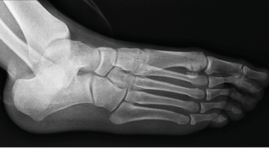
Figure 1. Oblique X-ray showing the bones of the foot.
The similar phylogenetic development of the human limbs suggests that the ontology of the lower limb may have many points in common with the development of the upper limb4.
Due to the fact that the foot demonstrates valid maturational parameters for use in legal studies and estimation of age5 and the use of a hand X-ray for bone assessment in the first two years of life has a series of limitations, the foot can be useful to complete the study of certain individuals, especially those who, due to special circumstances, cannot be subjected to radiographic examinations of the wrist (due to their absence, agenesis or hypoplasia derived from growth) or teeth (due to the lack of most of teeth)6.
The studies found in the scientific literature that use the foot as a structure endowed with interest in age prediction, do not contemplate the entire process of human growth, delimiting the conclusions in most cases at very early or very advanced ages, thus disregarding a wide age range that we consider may be useful in daily clinical practice2-4.
Bone maturation is directly influenced by intrinsic factors such as the race and sex of each individual, as well as by socioeconomic factors or nutrition7. The skeleton of the female sex ossifies before the male, in addition the ossification occurs following a symmetrical development and individual variations in the ossification of hereditary origin and character can occur8. Therefore, the estimation of bone age implies the observation of morphological features in the skeleton, as well as the comparison of the findings obtained on recent populations with known ages similar to those of the subject subject to study5.
The study of bone maturation to establish the age in individuals subject to clinical diagnosis, consists of a multitude of investigations aimed at achieving new methods that facilitate radiographic reading and that accurately approximate the assessment of bone age. Most of the methods published in the scientific literature refer to the radiological study of the anatomical region of the carpus. The use of metric or computerized methods are exposed to the scientific community as an alternative to facilitate clinical work. However, they are not considered first-choice methods, like X-rays of half the skeleton, discarded due to the experience required of the observer and the excess radiation absorbed by the patient9-10.
Currently, the most widely used methods for the study of bone maturation are divided into two large groups: qualitative methods, represented by the GP atlas11, and scoring methods, whose fundamental reference lies in the TW atlas, out on radiographs of the carpus12.
In addition, many of them are old, have a small sample, are considered on a population very different from the Caucasian population (reference population in the Spanish territory)13,14 or do not contemplate all the bones of the foot6, which allows us to propose a new investigation that improve these premises and consequently allow valid and updated conclusions to be obtained15.
For all these reasons, we believe that this research project can contribute interesting data to scientific knowledge and clinical utility in the determination of data and age prediction.
Marking us as objectives the quantification of the ossification ages of each foot bone and determine if there is a relationship between the ossification of the bones and the sex of the individual.
Patients and methods
Type of study and sample size
As described by Argimón16, the present study is cross-sectional, descriptive, observational and retrospective.
The study population was made up of 2476 digital radiographs, belonging to a total of 816 subjects in the growth period, which means an average of 3.04 radiographic projections per individual.
The x-rays consist of dorso-plantar load-bearing, lateral load-bearing and oblique projections of both right and left feet made by the Radiodiagnosis Services of the (SES), which represents all the images stored by the SES in the time interval included. between January 2007 and February 2011, of subjects with ages ranging from birth to 21 years. This means that the study includes the entire population of Extremadura in a period of growth that underwent radiological examination of the foot between 2007 and 2011.
The digital images were extracted directly from the PACS, or unique repository of digital images of the Autonomous Community of Extremadura, partially anonymized.
Inclusion criteria
After receiving the digital radiographs from the SES, they were selected and excluded those belonging to subjects whose characteristics could bias the final results of the study.
All the radiographs were performed on subjects undergoing radiological examination for different reasons, most of them traumatic, in which there were no lesions that affected bone structures (mainly fractures). Therefore, subjects who have been irradiated for reasons such as: growth studies, surgical interventions (pre- or post-surgical X-rays) and obvious structural, functional or traumatic alterations that affect bone morphology are considered reasons for exclusion from the sample.
Likewise, those radiographs that presented radiological artifacts were excluded from the study, were carried out unloaded, with doubtful visibility or technical failures derived from an incorrect position of the foot when performing the radiograph, in order to minimize the possible insecurity resulting from inter and intra explorer doubt when looking at the radiographs.
Equipment
The images that constitute the study sample are extracted from the only medical image repository that exists in the Autonomous Community of Extremadura. The PACS is centralized in the city of Mérida in the facilities of the Subdirectorate of Systems of the SES where the union point of all the radiodiagnostic Units of the Community is located.
In order to carry out this study and in accordance with its descriptive characteristics, it was considered necessary to start with a large sample that could be considered a population reference for the Autonomous Community of Extremadura. For this, a formal request is made to the SES, since it is the only Organization in the entire Autonomous Community that has a unique repository of digital medical images where all the radiographic studies carried out in the Public Health of Extremadura are stored.
Once the radiological studies were downloaded, they were recorded on DVD-type media and sent to the University Center of Plasencia UEX (Universidad de Extremadura) accompanied by DICOM-type software to be able to view them on any computer, after installing the program.
Once the samples were received, they were selected by applying the inclusion and exclusion criteria explained above. Once the valid extract was obtained, the radiographs were analyzed using the method described below and accumulating the study variables in an Excel spreadsheet that would allow their subsequent statistical analysis.
The analysis of the images was carried out by applying the method validated by Whitaker and designed to estimate the age in months on the X-ray of the foot skeleton14.
The method consists of applying a graduated scale of ossification stages for each of the bones that make up the growing foot. The system has three independent scales applicable to each bone of the foot, as long as it is in the formation process: scale to assess the degree of maturation of the first ossification nucleus, scale to assess the degree of maturation of the second ossification nucleus, and scale for assess the degree of union between them.
Viewing and analysis of the radiographs were carried out by two different investigators who would view the same radiographs at different time intervals in groups of 20 projections per session. These people are part of the teaching and research staff of the University of Extremadura UEX, professors of the Degree in Podiatry of the University Center of Plasencia and care podiatrists of the University Podiatry Clinic of the UEX.
In addition, analyzes of the series of 20 radiographs were viewed by the same person three times, separated by a period of 5 days.
Statistic analysis
The data have been analyzed with the software package SPSS version 19.0 for Windows.
The age and sex of the study population were described. The descriptive analysis of each study variable was provided, consisting of: ossification degree scale, number of cases, corresponding mean age of ossification expressed in months, standard deviation, 95% confidence interval, and lower and upper intervals expressed in months. In addition, contingency tables were made where the frequencies and percentages of each degree of ossification or maturational stage of the bone were shown with respect to the study population. All of the above, necessary to achieve the number one objective of this study consisting of quantifying and updating the ossification ages of each foot bone.
Results
Systematic review vs. current study in ossification of the bone
of the foot
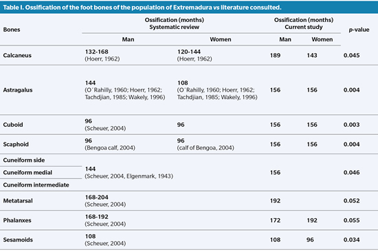
In Table I we can see the total ossification of the bones that comprise the foot, comparing the Extremaduran population against what the consulted literature dictates.
Contrast of ossification ages with respect to sex
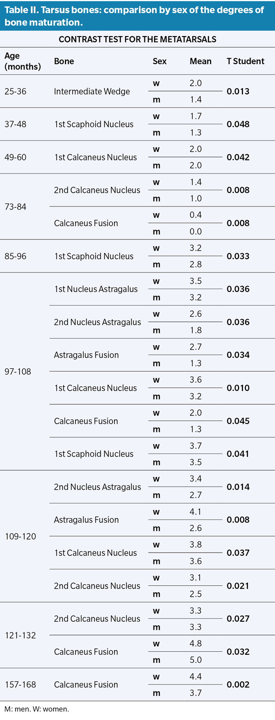
The data contrast study between the ages of ossification in girls and boys is carried out by applying the Student’s P Distribution for paired samples and the Levene Test of variance homogeneity, to verify the hypothesis of equality of variances between both sexes.
All the bones that make up the Tarsus have statistical significance in the comparison by sex with the exception of the Cuboid, the medial Cuneiform and the lateral Cuneiform. The rest of the tarsus bones have an explicit difference (P value ≤0.05) that translates into a discrepancy between the degree of ossification and its speed, later and slower for males (Table II).
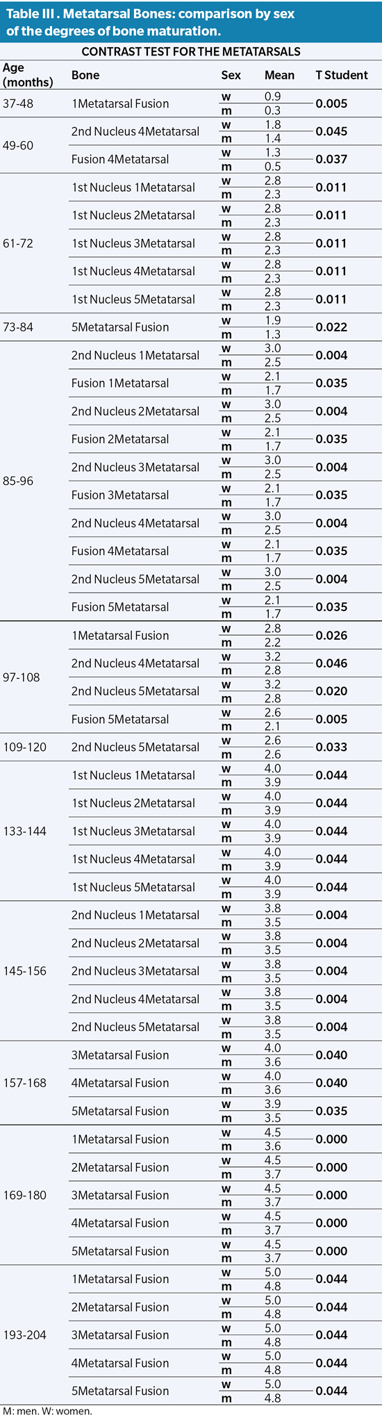
Similarly, the study of the metatarsals shows a delay in ossification of boys with respect to girls. All the ossification nuclei of the Metatarsus are significant, however, it should be noted that the first ossification nucleus of the Metatarsals has a difference by sex greater than 5 to 6 years (61 to 72 months with P value ≤0.011). The same occurs with the ossification of the second metatarsal ossicle between 7 and 8 years (85 to 96 months with P value ≤0.004). The fusions between primary and secondary nuclei have their greatest statistical significance around the mean age interval of 14 to 15 years (169-180 months with P value ≤0.000), where the delay in the rate of ossification in sex becomes more evident. male versus female (Table III).
Regarding the results obtained regarding the differences in ossification between both sexes for the finger bones, it should be noted that all the Phalanges are significant, showing a delay in the ossification of the male sex with respect to the female sex (Table IV).
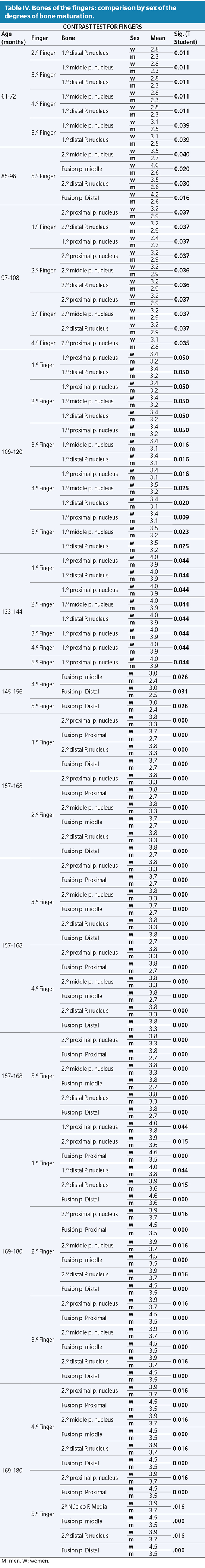
However, it is worth noting the statistical significance for the first ossification nuclei of both the middle and distal Phalanges, from the mean age interval of 5 to 6 years (61 to 72 months with P value ≤0.011). For the first ossification nuclei of the proximal phalanges from 9 to 10 years of age (109 to 120 months with P value ≤ 0.050) (Table I).
The second ossification nuclei of the Phalanges, as well as the fusions between them, have a slower ossification rate for males compared to females, very clearly in the mean age interval between 13 and 14 years (157 to 168 months with P value ≤ 0.000) (Table I).
Discussion
Our study is the only one, to date, that includes the entire population of the Autonomous Community of Extremadura subjected to a radiological study of the foot and ankle, in individuals in the growth period. For this reason throughout the writing of this work the term “sample” is not included, considering instead the term “population”.
It should be noted that most of the authors with relevant scientific publications in this field insist on the differences implied by the study of bone maturation in different populations based on race, social or economic level 8 we consider it remarkable to have in this study all the the Community of Extremadura, representing the Spanish population and in its maximum extrapolation to the Caucasian race17.
Bearing in mind that the results obtained in a certain race are not applicable to another, those studies published in reference to other populations other than the Spanish or to other races other than the Caucasian are discarded for the writing of this point. This premise leaves us few opportunities to compare our results, since the foot is a structure little studied in the field of bone maturation and age determination15.
In general terms, the results obtained after the complex study of bone maturation in the bones of the foot, coincide in most cases with the scientific publications on the subject.
However, if we analyze the foot bones one by one, we can see how there are variations between the published ages with respect to our results. These differences, minimal in some cases, do not fail to provide additional information about the bone, which makes it possible to further specify the estimation of bone data. Bearing in mind that there are certain situations in which the accuracy in the estimation of age is extremely important (clinical diagnoses in growth disorders, legal links, estimation of the data of skeletal remains or judicial processes, among others), we consider it highly It is of interest to be able to specify as much as possible the estimate of the age of an individual9,11,16.
Analyzing in depth the process of ossification of each one of the bones of the foot, we can begin by affirming that among the bones that form the Tarsus there are significant differences between what has been published by various authors and our results4. Regarding the Calcaneus, for example, we differ in our results regarding epiphyseal fusion at term, since our results establish it from 180 months (some 9 months later in boys than in girls), compared to ages earliest proposed by the other authors18. The talus has a bone maturation that is more similar to that established by the classical bibliography. However, complete epiphyseal fusion in the literature shows ages of 108 months in children, while in the population of Extremadura it is 156 months with 108 months and 167 months for females and males respectively19,20.
On the other hand, it is worth noting the age of total maturation of the Cuboid between 72 and 108 months in girls, compared to 60 to 96 months in boys, compared to the 96 months of age established by Seucher without differentiation by sex. This fact also happens with the scaphoid4,21.
The values obtained after our analysis in the metatarsals do not differ much from the ages established in the scientific literature, however, it is worth noting the simultaneous maturational behavior among the five metatarsals, which differs with some authors who affirm that the first metatarsal ossifies. regardless of the rest22. Something similar occurs with the study of fingers. Some authors affirm the difference in the maturational rhythm between the Phalanges of the different fingers, as well as between themselves4,18 a matter in which we disagree, since our results show, in general lines, a uniform maturational rhythm in the entirety of the fingers and between Phalanges23.
The method used by Whitaker was interesting due to the almost complete reduction of inter- and intra-observer bias (very frequent in observational methods), in addition to considering the age of the study individuals in months, which specifies the results making them more exact14. The only weakness of the method is that it does not include all the bones of the foot for study, so we decided to apply this method, scientifically validated, to all the ossification nuclei of the foot and ankle bones (primary, secondary and mergers between the two) in order to expand their initial study by providing predictive data models for the skeleton of the foot and ankle15,18.
Our results coincide with what is established in the classical literature regarding the differentiation of bone maturation by sex, confirming that epiphyseal fusions occur earlier in females than in males. In addition, we affirm that this situation occurs not only in bone fusions but also in the appearance of most of the nuclei, both primary and secondary, according to what was established by Seucher4.
Nor is the rate of maturation the same for both sexes. This issue was already mentioned by Robledo without specifying exactly which of the two sexes evolved faster or slower. Based on our results, we are able to affirm that the rate of maturation is slower in boys than in girls6.
Based on the results obtained and in relation to the objective set forth in this investigation, it is concluded that, with respect to the variability of the normal course of the ossification process between sexes, we affirm that there are differences that imply slower bone maturation for the males compared to females, and regarding the ossification of the foot bones, the population of Extremadura has a later total ossification than the population reported in the literature14-17,22.
Ethical considerations of the study
As it is not a study related to human health that involves invasive procedures or direct intervention with patients, approval by any Ethical Committee for Experimentation or Bioethics was not necessary in accordance with the provisions of Article 1 of Law 14/2007. of July 3 of Biomedical Research.
The researchers and health professionals related to this study undertook to ensure compliance with Organic Law 15/1999 of December 13 on the Protection of Personal Data and Royal Decree 1720/2007 of December 21, which approves the development regulations of the LOPD.
The clinical data handled in this research have been treated with the utmost confidentiality and custody of the information in accordance with the provisions of Law 41/2002 of November 14, Basic Regulation of Patient Autonomy.
Conflict of interest
There is no conflict of interest on the part of the authors.
Funding
None.
References
- Garamendi PM, Landa MI, Ballesteros J, Solano MA. Reliability of the methods applied to assess age minority in living subjects around 18 years old. A survey on a Moroccan origin population. Forensic Sci Int. 2005;154(1):3-12. DOI: 10.1016/j.forsciint.2004.08.018.
- Gisbert Calabuig JA. Medicina Legal y Toxicología. Barcelona: Masson S.A.; 2004.
- Krogman WM, Isçan MY. The Human eskeleton in Forensic Medicine. Springfield: Charles C Thomas; 1986.
- Scheuer L, Black SM. The Juvenile Skeleton. San Diego: Elsevier Academic Press; 2004.
- Robledo-Acinas MM. Determinación de la edad ósea en adolescentes. Estudio radiológico del pie y análisis de imágen en el grupo de 17 a 19 años [Tesis Doctoral]. Departamento de Toxicología y Legislación sanitaria. Madrid; Universidad Complutense de Madrid; 2008.
- Tanner JH, Whitehouse RM. Standard skeletal maturity. Part I. París: International Children’s Center; 1959.
- Reverte JM. Antropología Forense. Madrid: Ministerio de Justicia; Secretaría General Técnica. Centro de Publicaciones; 1990.
- Scheuer L, Black SM. Developmental Juvenile Osteology. San Diego, CA: Academic Press; 2000.
- Argemí J, López-Cuevas I. Nuevo método para el estudio de la maduración ósea por ordenador. An Esp Pediatr. 1987;27(30):85-8.
- Ebritorn B, Altarriba J. Presentación de un nuevo método biométrico (TVO) para la valoración de la edad ósea en niños. Med Kin. 1979;214:50-6.
- Greulich WW, Pyle SL. Radiographic atlas of skeletal development of the hand and wrist. California: Standford University Press; 1959. DOI: 10.1097/00000441-195909000-00030.
- Tanner JM, Whitehouse RM. Assessment of skeletal maturity and presiction of adult height (TW2). London: London Academic Press; 1993.
- Hernandez C, Sánchez E, Sobradillo B, Rincón JM, Narvaiza JL.. A new method for assessment of skeletal maturity in the first 2 years of life. Pediatr Radiol. 1988;18:484-9. DOI: 10.1007/BF00974086.
- Whitaker JM, Rousseau L, Williams T, Rowan RA, Hartwig WC. Scoring system for estimating age in the foot skeleton. Am J Phys Anthropol. 2002;118(4):385-92. DOI: 10.1002/ajpa.10109.
- Crowder C, Austin D. Age ranges of epiphyseal fusion in the distal tibia and fibula of contemporary males and females. J Forensic Sci. 2005;50(5):1001-7. DOI: 10.1520/JFS2004542.
- Argimon Pallés JM, Jiménez Villa J. Métodos de investigación clínica y epidemiológica. 4.ª ed. Madrid: Elsevier; 2013.
- Krogman WM, Isçan MY. The Human eskeleton in Forensic Medicine. Springfield: Charles C. Thomas; 1986.
- Hoerr NL, Pyle SL, Francis CC. Radiographic Atlas of Skeletal Development of the Foot and Ankle. A Standard of Reference. Springfield: C.C. Thomas; 1962.
- O´Rahilly R, Gardner E. The skeletal development of the foot. Clinical Orthopaedics and Related Research. 1960;16:7-14.
- Tachdjian MO.The Child´s Foot. Philadelphia: W.B. Saunders Company; 1985.
- Wakely CJ, Johnson DP, Watt I. The value of MR imaging in the diagnosis of the os trigonum syndrome. Skeletal Radiology. 1996;25(2):133-6. DOI: 10.1007/s002560050049.
- Franch M, Infante MM. Cronología de osificación del pie. Radiogonometría. El Peu. 2004;24(3):148-58.
- Birkner R . Normal Radiographic Patterns and Variances of the Human Skeleton-An X Ray Atlas of Adults and Children. Baltimore (Munich): Urban and Schwanzemberg; 1978.